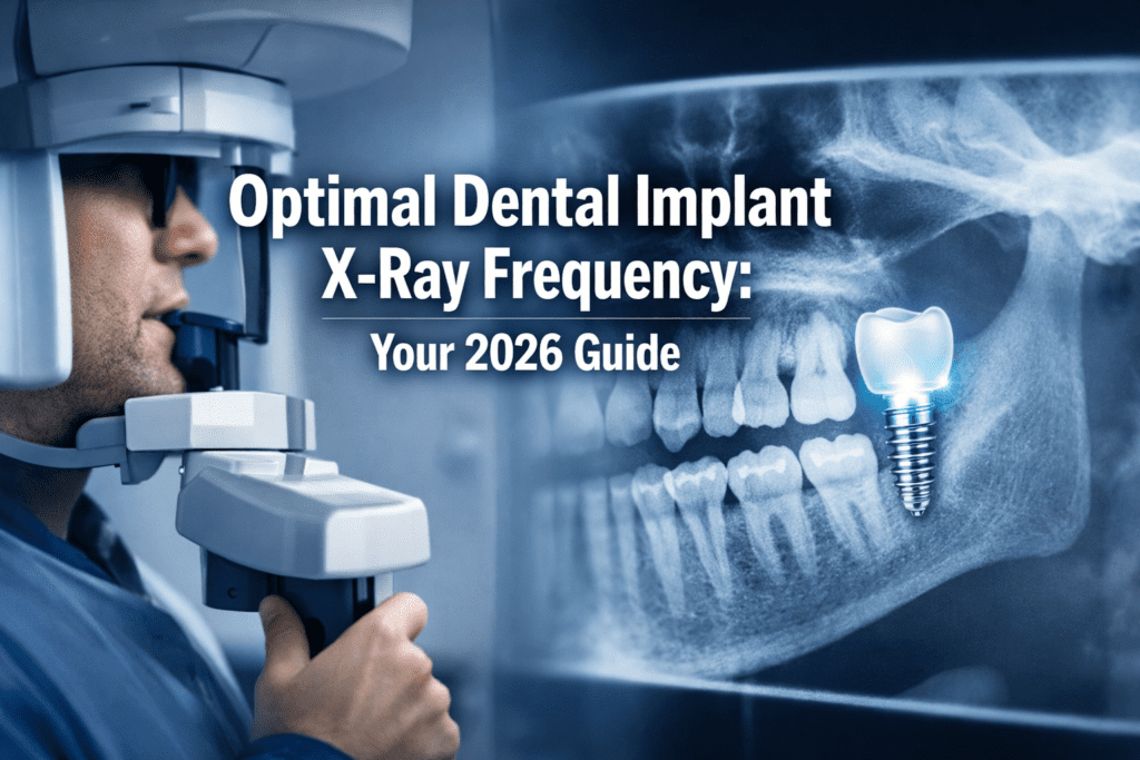
Embarking on the journey of dental implants is an investment in your oral health and overall well-being. These modern marvels of dentistry offer a durable, aesthetic, and functional solution for missing teeth. However, like any significant investment, they require careful monitoring to ensure their longevity and success. A critical aspect of this ongoing care is determining the appropriate dental implant x-ray frequency. Understanding why, when, and how often X-rays are taken for your dental implants is paramount for both patients and practitioners, especially in 2026, as technology and best practices continue to evolve. This comprehensive guide will illuminate the crucial role of dental imaging in implant maintenance, helping you feel confident and informed about your treatment.
Key Takeaways
- X-rays are essential for monitoring implant health: They allow dentists to visualize bone integration, detect complications like peri-implantitis early, and assess the implant’s structural integrity.
- Frequency varies greatly: The ideal dental implant x-ray frequency is highly individualized, depending on the implant stage (pre-placement, post-surgery, long-term maintenance), patient health, and risk factors.
- Initial X-rays are crucial for planning and immediate post-op: Multiple X-rays are typically taken during the planning and placement phases to ensure precise positioning and confirm initial success.
- Regular follow-ups are non-negotiable: Even healthy implants require periodic X-rays (e.g., annually or biennially) for long-term monitoring and proactive problem-solving.
- Technological advancements minimize radiation: Modern digital X-rays and 3D imaging offer high diagnostic value with significantly reduced radiation exposure, making them safer and more efficient.
Why X-Rays are Indispensable for Dental Implants
Dental implants are unique in that they are surgically placed directly into your jawbone, acting as artificial tooth roots. Unlike natural teeth, which have a periodontal ligament for shock absorption and sensory feedback, implants fuse directly with the bone through a process called osseointegration. This intimate connection means that conventional visual examinations alone aren’t enough to assess their health. This is precisely where X-rays become indispensable.
X-rays provide a two-dimensional (and sometimes three-dimensional) view of structures that are otherwise hidden beneath the gums and bone. They allow your dental professional to:
- Assess Bone Quality and Quantity: Before implant placement, X-rays are vital for evaluating the existing bone to determine if it can adequately support an implant. This includes identifying areas that might require a bone graft recovery dental implant to ensure sufficient bone volume.
- Confirm Osseointegration: After the implant is placed, X-rays are used to monitor the healing process and confirm that the implant is successfully fusing with the jawbone. This bone-to-implant contact is crucial for stability.
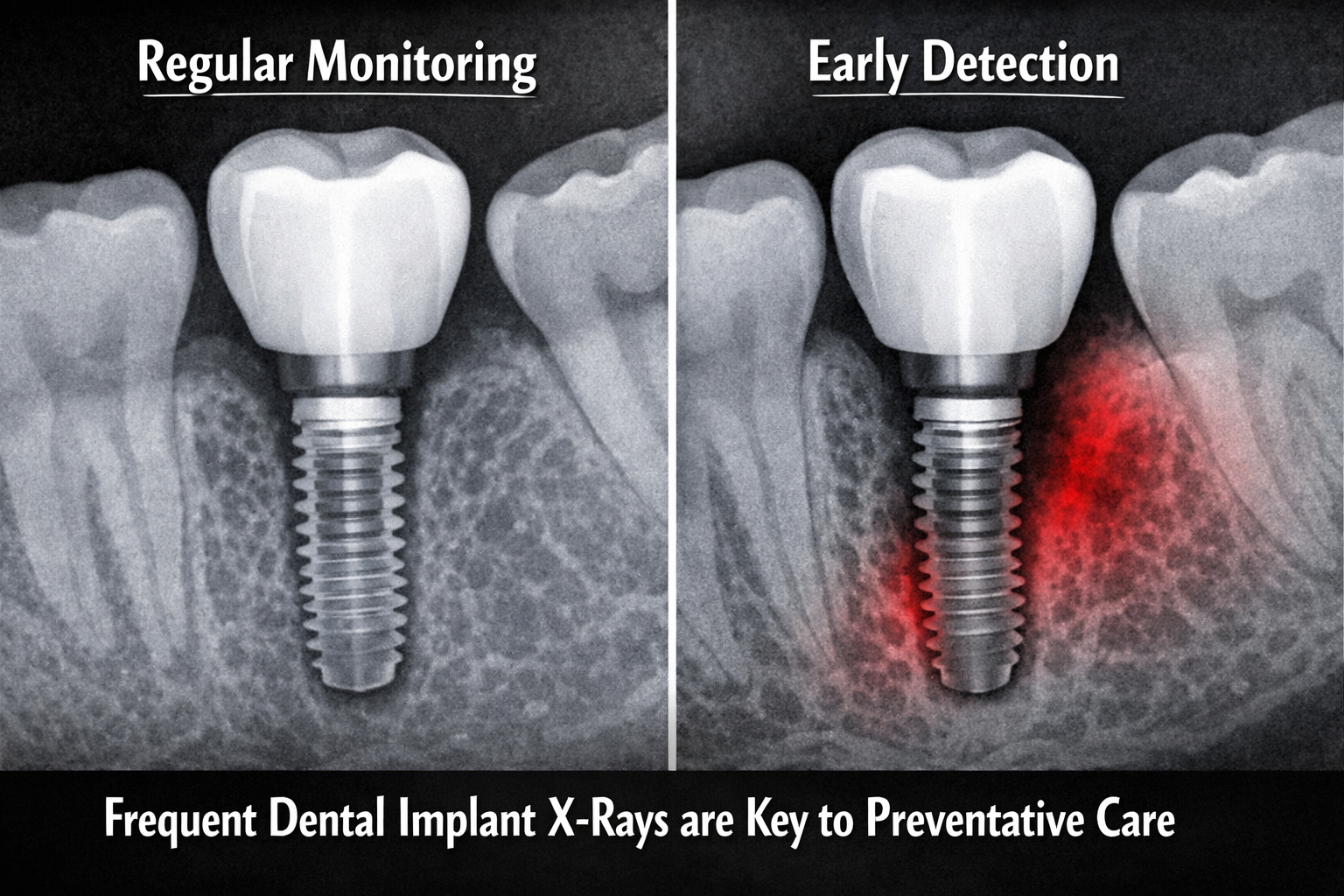
- Detect Early Complications: One of the most significant benefits of X-rays is their ability to detect problems early, often before they become symptomatic. These can include:
- Peri-implantitis: An inflammatory condition affecting the tissues surrounding a dental implant, leading to progressive bone loss. Early detection through X-rays allows for timely intervention, potentially saving the implant.
- Loosening of Components: While rare, components like the abutment or crown can sometimes loosen. X-rays can help identify such issues.
- Fractures: Although implants are incredibly strong, X-rays can reveal hairline fractures in the implant itself or the surrounding bone.
- Infection: Persistent localized infections around the implant can be identified.
- Monitor Surrounding Structures: X-rays also help ensure the implant isn’t encroaching on vital structures like nerves, sinuses, or adjacent teeth.
- Evaluate Long-Term Stability: Over the years, routine X-rays serve as a baseline for comparison, allowing your dentist to track any subtle changes in bone level or implant position.
Without the detailed internal view provided by X-rays, many potential issues would go unnoticed until they become advanced and more challenging (and expensive) to treat. Therefore, understanding the rationale behind your dental implant x-ray frequency is the first step in proactive implant care.
Initial Stages: Planning and Placement X-Ray Frequency
The journey of dental implant placement is carefully orchestrated, and X-rays play a starring role in almost every act. The dental implant x-ray frequency during the initial stages is typically higher than during long-term maintenance, reflecting the critical need for precision and immediate post-operative assessment.
Pre-Surgical Planning
Before a single incision is made, extensive imaging is performed. This phase is crucial for ensuring optimal placement and avoiding complications.
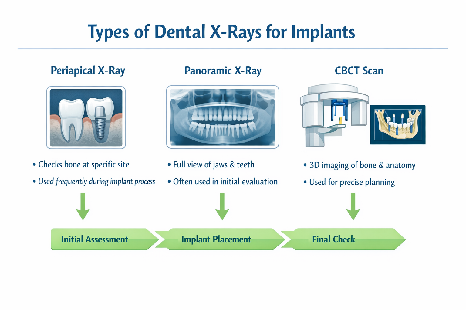
- Panoramic X-ray: Often the first diagnostic image, a panoramic X-ray captures a broad view of your entire mouth, including both jaws, sinuses, and temporomandibular joints. It helps assess overall bone structure, identify nerve locations, and detect any underlying pathologies.
- Periapical X-rays (PAs): These detailed X-rays focus on specific teeth or areas, providing a very clear view of a few teeth from crown to root, including the surrounding bone. They are excellent for assessing bone height and width at the proposed implant site.
- Cone Beam Computed Tomography (CBCT): This is perhaps the most revolutionary imaging tool for implant dentistry. A CBCT scan provides a 3D view of the jawbone, offering unprecedented detail on bone quality, density, and the exact relationship to vital structures like the inferior alveolar nerve or maxillary sinus. This 3D data allows for virtual implant planning, precise measurement of bone dimensions, and identification of areas where additional procedures like sinus lifts or bone grafting may be necessary. For complex cases or full-mouth restorations like all-on-4 dental implants Dallas, a CBCT is almost always standard.
Typical Pre-Surgical X-Ray Frequency:
Multiple types of X-rays will be taken as needed during this phase. It’s not uncommon to have a panoramic, several periapical, and a CBCT scan within a few weeks or months leading up to surgery. The specific number depends on the complexity of the case and the information required by the surgeon.
During and Immediately Post-Surgical Placement
Once the implant surgery is underway, X-rays continue to be vital.
- Intra-Operative X-rays: Sometimes, an X-ray (usually a periapical) might be taken during the surgery itself to confirm the precise angulation and depth of the implant placement before final seating. This ensures the implant is perfectly positioned.
- Immediate Post-Operative X-rays: Right after the implant is placed, a periapical X-ray is almost always taken. This image serves several critical purposes:
- Confirmation of Placement: It verifies that the implant is in the correct anatomical position.
- Baseline for Future Comparison: This initial X-ray acts as a crucial baseline. Future X-rays will be compared to this one to monitor bone levels and detect any changes over time.
- Detection of Immediate Issues: It can identify any unforeseen issues directly related to the placement, though these are rare with proper planning.
Typical Post-Surgical X-Ray Frequency:
At least one periapical X-ray is taken immediately post-surgery. Depending on the complexity, another might be taken during the initial healing period (e.g., 2-4 weeks later) to assess early bone healing, especially if there were any concerns during surgery or if a bone graft was performed. Learn more about the complete dental implant recovery timeline.
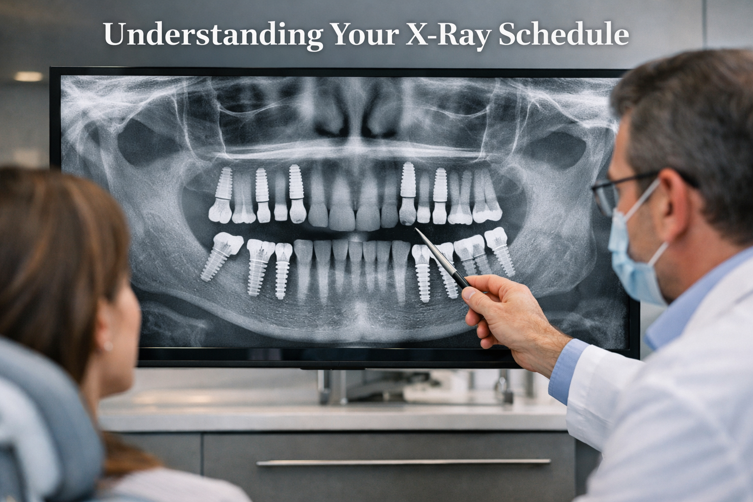
Long-Term Maintenance: The Evolving Dental Implant X-Ray Frequency
Once your dental implant has successfully integrated and the final crown or bridge has been placed, the focus shifts to long-term maintenance. This phase is where the discussion around dental implant x-ray frequency becomes more nuanced and patient-specific. The goal is to monitor the implant’s health and the surrounding bone without unnecessary radiation exposure.
Establishing a Baseline and Regular Check-ups
After the final restoration (crown, bridge, or denture) is securely attached to your implant, another X-ray is typically taken. This X-ray establishes the definitive “baseline” for your fully restored implant. This is an extremely important image, as it will be used for comparison for many years to come.
- First Year Post-Restoration: During the first year after your final restoration, your dentist might recommend an X-ray (usually a periapical) at your 6-month or 1-year check-up. This allows them to confirm continued stability and integration and ensure there are no early complications with the prosthetic components.
- Annual to Biennial Check-ups: For most healthy, stable dental implants, the general recommendation for long-term monitoring involves X-rays every one to two years [1]. This frequency allows your dentist to:
- Monitor Bone Levels: The primary concern is detecting any changes in the bone level around the implant, which could indicate peri-implantitis or other issues.
- Assess Prosthetic Integrity: Check for any signs of loosening or fracture of the crown, abutment, or even the implant itself.
- Evaluate Surrounding Dentition: Ensure the implant is not negatively affecting adjacent teeth or vice-versa.
Quote:
“Regular, targeted X-rays are the silent guardians of your dental implant’s health. They allow us to see what the naked eye cannot, empowering early intervention and significantly extending the lifespan of your investment.”
— Dr. Jane Doe, Implantologist
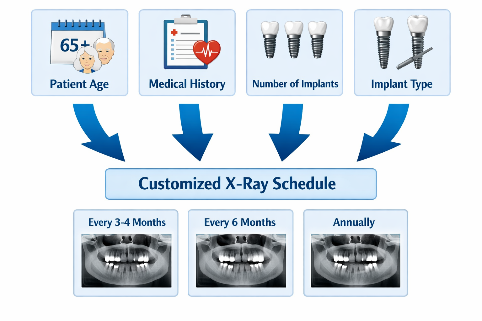
Factors Influencing Dental Implant X-Ray Frequency
While annual to biennial check-ups are a general guideline, several factors can alter the recommended dental implant x-ray frequency:
Patient’s Overall Health:
- Systemic Diseases: Conditions like uncontrolled diabetes, osteoporosis, or certain autoimmune diseases can increase the risk of implant complications and may warrant more frequent imaging [2].
- Smoking: Smokers have a significantly higher risk of peri-implantitis and bone loss, often requiring more vigilant monitoring.
- Medications: Certain medications, especially those affecting bone metabolism (e.g., bisphosphonates), can influence implant health and the need for closer observation.
Implant-Specific Factors:
- Number of Implants: Patients with multiple implants, especially full-arch restorations like all-on-6 full-arch implants, may have different imaging needs compared to those with a single implant.
- Implant Location: Implants in areas of higher chewing forces or those closer to vital anatomical structures might require more frequent checks.
- Implant History: An implant that has previously shown minor issues or required intervention (e.g., treatment for early peri-implantitis) will likely be monitored more closely.
Oral Hygiene Practices:
- Patients with excellent oral hygiene and no signs of gum disease or inflammation around their implants might require less frequent X-rays.
- Conversely, those with suboptimal hygiene or a history of periodontal disease (which can contribute to peri-implantitis) may need more frequent imaging.
Clinical Findings:
- If your dentist notices any clinical signs during a routine exam (e.g., redness, swelling, bleeding upon probing, or changes in gum tissue around the implant), an X-ray will immediately be taken regardless of the scheduled frequency to investigate further.
Table: General Guidelines for Dental Implant X-Ray Frequency (2026)
| Stage of Implant Treatment | Type of X-ray(s) Commonly Used | Typical Frequency | Purpose |
|---|---|---|---|
| Planning & Pre-Surgery | Panoramic, Periapical, CBCT | As needed (multiple) | Assess bone, vital structures, plan placement. |
| Post-Surgery (Immediate) | Periapical | Once, immediately after placement | Confirm position, establish baseline. |
| Healing Phase | Periapical | 1-3 months post-op (if needed) | Assess early healing, bone integration. |
| Abutment Placement | Periapical | At time of placement (often) | Confirm abutment seating. |
| Post-Restoration | Periapical | Once, after final crown/bridge | Establish final baseline. |
| Long-Term Maintenance (Healthy Implant) | Periapical | Every 1-2 years [1] | Monitor bone levels, detect subtle changes. |
| Long-Term Maintenance (High-Risk/Complication) | Periapical, potentially CBCT | Every 6-12 months, or as clinically indicated | Close monitoring for peri-implantitis, bone loss. |
(Note: These are general guidelines. Your dentist will determine the precise schedule based on your individual needs.)
Understanding X-Ray Types and Safety for Dental Implants

When discussing dental implant x-ray frequency, it’s also important to understand the different types of X-rays used and the safety measures in place. Advancements in dental radiography have significantly reduced radiation exposure, making these diagnostic tools safer than ever.
Common X-Ray Types for Implants
Periapical (PA) X-rays:
- What they show: A very detailed view of one or two teeth, from the crown to the root and the surrounding bone. Ideal for assessing the bone level directly adjacent to the implant.
- Use in implants: Crucial for initial baseline, post-operative checks, and long-term monitoring of bone levels around individual implants.
Panoramic X-rays:
- What they show: A wide view of the entire mouth, including all teeth, both jaws, sinuses, and temporomandibular joints.
- Use in implants: Useful for initial screening, assessing overall bone structure, and identifying larger anatomical landmarks. Less detailed for individual implant monitoring compared to PAs.
Cone Beam Computed Tomography (CBCT):
- What they show: A 3D image of the mouth and facial structures. Provides detailed cross-sectional views of bone, soft tissues, and nerve pathways.
- Use in implants: Invaluable for complex treatment planning, determining precise implant placement, assessing bone volume for grafting, and diagnosing complex issues like extensive peri-implantitis or nerve involvement. While a CBCT involves more radiation than a 2D X-ray, its diagnostic yield is significantly higher for certain situations, making it a necessary tool in specific circumstances. Learn more about CT scan and dental implants.
Radiation Safety and Modern Technology
One common concern patients have about repeated X-rays is radiation exposure. It’s important to recognize that we are exposed to background radiation from natural sources every day. Dental X-rays contribute a very small amount to this total.
- Digital Radiography: The vast majority of dental practices today use digital X-ray systems. These systems have drastically reduced radiation exposure compared to traditional film X-rays—by as much as 80-90% [3]. Digital images also appear instantly, can be enhanced, and are environmentally friendly as they don’t require chemical processing.
- Lead Aprons and Collars: You will always be provided with a lead apron and often a thyroid collar to shield the rest of your body from any stray radiation.
- Targeted Imaging: Dentists are trained to take only the necessary X-rays. The principle of “as low as reasonably achievable” (ALARA) is strictly followed, meaning the lowest possible radiation dose is used to obtain diagnostically acceptable images.
- Benefit-Risk Assessment: Your dental professional always weighs the diagnostic benefit of an X-ray against the minimal risk of radiation exposure. For dental implants, the benefit of early detection of complications often far outweighs the minimal risk.
When considering your dental implant x-ray frequency, trust that your dental team is adhering to strict safety protocols and utilizing the most advanced technology to protect your health while ensuring the long-term success of your implants. Feel free to discuss any concerns you have about radiation with your dentist. For more information about our practice, visit our About Us page.
The Role of the Patient and Dentist in Determining X-Ray Frequency
Optimizing your dental implant x-ray frequency is a collaborative effort between you, the patient, and your dental team. While guidelines exist, the final decision is always personalized, taking into account a holistic view of your oral and general health.
Your Role as a Patient
As a patient, your active participation is invaluable in determining the appropriate X-ray schedule.
- Maintain Excellent Oral Hygiene: This is perhaps the most crucial factor within your control. Meticulous brushing, flossing, and using appropriate interdental cleaners (like water flossers or interdental brushes recommended for implants) are essential for preventing peri-implantitis. Patients with impeccable oral hygiene and no signs of inflammation may require less frequent X-rays. Learn about the best dental implant aftercare products.
- Attend Regular Dental Check-ups: Consistency is key. Even if your implants feel fine, attending your scheduled recall appointments allows your dentist to perform visual examinations, probing, and professional cleanings, which are vital for implant maintenance. These check-ups are when the need for X-rays is often assessed.
- Report Any Concerns Immediately: If you experience any symptoms such as pain, swelling, bleeding, a loose feeling around the implant, or difficulty chewing, contact your dentist right away. These symptoms would trigger an immediate X-ray, regardless of your last one.
- Provide Accurate Medical History: Inform your dentist about any changes to your medical history, medications you are taking, or lifestyle habits (like smoking). These factors directly influence your risk profile and, consequently, your recommended X-ray frequency.
- Ask Questions: Don’t hesitate to ask your dentist why an X-ray is being recommended or what they are looking for. An informed patient is an empowered patient.
Your Dentist’s Role
Your dentist is the expert guiding your implant care. Their role in determining dental implant x-ray frequency involves:
- Comprehensive Assessment: Combining your medical and dental history with clinical examination findings (visual inspection, probing depths, signs of inflammation).
- Risk Stratification: Evaluating your individual risk factors for implant complications (e.g., history of periodontitis, smoking, systemic diseases). Patients at higher risk will generally have a more frequent X-ray schedule.
- Adherence to Professional Guidelines: Following established guidelines from dental organizations (like the American Academy of Periodontology or American Dental Association) while tailoring them to your specific needs.
- Utilizing Advanced Technology: Employing modern digital X-ray systems to minimize radiation exposure while maximizing diagnostic yield.
- Patient Education: Clearly explaining the rationale behind the recommended X-ray schedule, what they are looking for, and what the X-ray results indicate.
- Collaborative Decision-Making: Engaging you in the decision-making process, ensuring you understand and are comfortable with the proposed maintenance plan.
Ultimately, your dental implant care plan, including the frequency of X-rays, is a dynamic one. It will adapt and evolve based on your implant’s health, your overall health status, and any new clinical findings. Trusting your dental team and being an active participant in your care are the best ways to ensure the long-term success and health of your dental implants. For complex cases or a second opinion on your implant needs, consider a dental implant consultation with our specialists.
Dental Implant X-Ray Frequency Advisor 🦷
Use this tool to get an estimated **dental implant x-ray frequency** based on common factors. *Always consult with your dental professional for personalized advice.*
Conclusion
Understanding your dental implant x-ray frequency is not merely about enduring a diagnostic procedure; it’s about actively participating in the long-term success of your dental implants. From the intricate planning stages where 3D CBCT scans precisely map your anatomy, through the critical post-surgical checks that confirm successful osseointegration, to the periodic maintenance X-rays that safeguard against unforeseen complications, imaging plays an irreplaceable role.
In 2026, with advanced digital radiography and the ALARA principle guiding every decision, the radiation exposure from dental X-rays is minimal, especially when weighed against the profound benefits of early detection and intervention. Your personalized X-ray schedule is a testament to the individualized care your dental team provides, factoring in your unique health profile, the specific characteristics of your implants, and your commitment to oral hygiene.
Actionable Next Steps:
- Discuss with Your Dentist: At your next dental appointment, ask your dentist about your specific dental implant x-ray frequency schedule and the rationale behind it.
- Maintain Oral Hygiene: Continue your diligent oral hygiene routine (brushing, flossing, interdental cleaning) to minimize the risk of complications.
- Attend Regular Check-ups: Do not skip your scheduled dental appointments. These are crucial for both visual and radiographic assessment.
- Report Any Changes: If you notice any discomfort, swelling, or changes around your implant, contact your dentist promptly. Early action can prevent minor issues from becoming major problems.
By staying informed and engaged, you empower yourself to protect your dental implant investment and enjoy a lifetime of healthy, confident smiles.
References
[1] American Academy of Periodontology. (2005). “Dental Implants: Patient Selection, Management, and Maintenance”. Journal of Periodontology, 76(8 Suppl), 1547-1555. (Note: Specific guidelines may be updated, consult the latest AAP recommendations.)
[2] Renvert, S., & Quirynen, M. (2019). “Risk factors for peri-implantitis.” Periodontology 2000, 81(1), 22-34.
[3] White, S. C., & Pharoah, M. J. (2018). Oral Radiology: Principles and Interpretation (8th ed.). Elsevier.
Leave a Reply
Share your thoughts or ask a question about dental implants. Your email address will not be published.