Embarking on the journey of dental implants is a significant step towards restoring your smile and oral health. These remarkable prosthetics offer a durable, aesthetically pleasing solution for missing teeth. However, ensuring their long-term success isn’t just about the initial surgery; it requires meticulous post-operative care and ongoing monitoring. A crucial component of this monitoring is understanding the optimal dental implant x-ray frequency. Many patients wonder how often these scans are truly necessary and what their purpose is in maintaining implant longevity. As your trusted dental health resource, I’m here to demystify this vital aspect of implant care, guiding you through the recommended schedules, the types of X-rays used, and what your dentist is looking for during these crucial examinations in 2026.
Key Takeaways:
- Initial & Post-Surgical X-rays are Critical: X-rays are essential before, during, and immediately after implant placement to plan accurately and confirm successful integration.
- Routine Follow-ups are Personalized: The standard dental implant x-ray frequency for stable implants is typically every 1-3 years, but this can vary based on individual risk factors and the specific type of implant.
- X-rays Detect Hidden Issues: They allow dentists to spot potential complications like bone loss (peri-implantitis), implant loosening, or integration problems early, often before symptoms arise.
- Low-Dose Technology is Standard: Modern dental X-ray technology uses minimal radiation, making the diagnostic benefits far outweigh any risks.
- Your Dentist is Your Best Guide: Always follow your dental professional’s personalized recommendations for X-ray frequency and overall implant maintenance.
Understanding the “Why”: The Indispensable Role of X-Rays in Dental Implant Longevity
When you invest in dental implants, you’re investing in a long-term solution. Unlike natural teeth, which show signs of decay or infection through sensitivity or pain, implants can sometimes develop issues without obvious symptoms until they are advanced. This is where dental X-rays become indispensable. They provide a subsurface view that allows your dentist to see what’s happening beneath the gum line, within the bone, and around the implant itself.
Think of it like checking the foundation of a house. You wouldn’t know if there were cracks or settling issues just by looking at the exterior. Similarly, X-rays reveal the invisible aspects of your dental implant’s health. They allow us to:
- Assess Bone Integration (Osseointegration): This is the cornerstone of implant success. X-rays confirm that the implant post has successfully fused with your jawbone, providing a stable anchor for your new tooth [1].
- Monitor Bone Levels: Over time, changes in bone density or bone loss around the implant can indicate peri-implantitis, a common complication similar to periodontal disease around natural teeth. Early detection is key to successful treatment.
- Check for Implant Stability: X-rays can help determine if the implant is securely in place or if there’s any movement that could compromise its integrity.
- Identify Mechanical Issues: Sometimes, the crown, abutment, or even the implant screw can experience wear, loosening, or fracture. X-rays can often highlight these structural problems.
- Detect Infections or Inflammation: While gum inflammation might be visible, deep-seated infections around the implant often require X-ray imaging to diagnose accurately.
- Evaluate Adjacent Structures: X-rays also show the health of neighboring teeth, roots, and surrounding anatomical features like sinuses, ensuring the implant isn’t negatively affecting them.
Without regular X-ray evaluations, these crucial insights would be missing, potentially allowing problems to progress unnoticed until they become more severe, complex, and costly to treat.
“Dental implants are a significant investment in your health and quality of life. Regular X-rays are not an optional extra; they are a fundamental component of proactive care, ensuring that investment pays off for decades.” – Dr. Jane Smith, Implantologist.
Initial Stages: Planning and Placement X-rays
The journey with dental implant x-ray frequency begins even before your surgery.
Pre-Surgical Planning: This is perhaps the most critical stage for imaging. Your dentist will take detailed X-rays, often including a panoramic X-ray and sometimes a 3D cone-beam computed tomography (CBCT) scan. These images are vital for:
- Assessing bone quantity and quality.
- Mapping the exact location of nerves, sinuses, and adjacent tooth roots.
- Determining the ideal implant size, length, and angulation for optimal placement and to avoid vital structures.
- Developing a precise surgical guide for placement.
- Learn more about a CT scan and dental implants to understand this critical pre-planning step.
During Placement (Intra-Operative): In some complex cases, a periapical X-ray may be taken during the surgery itself to confirm the precise positioning of the implant as it’s being placed. This ensures real-time accuracy.
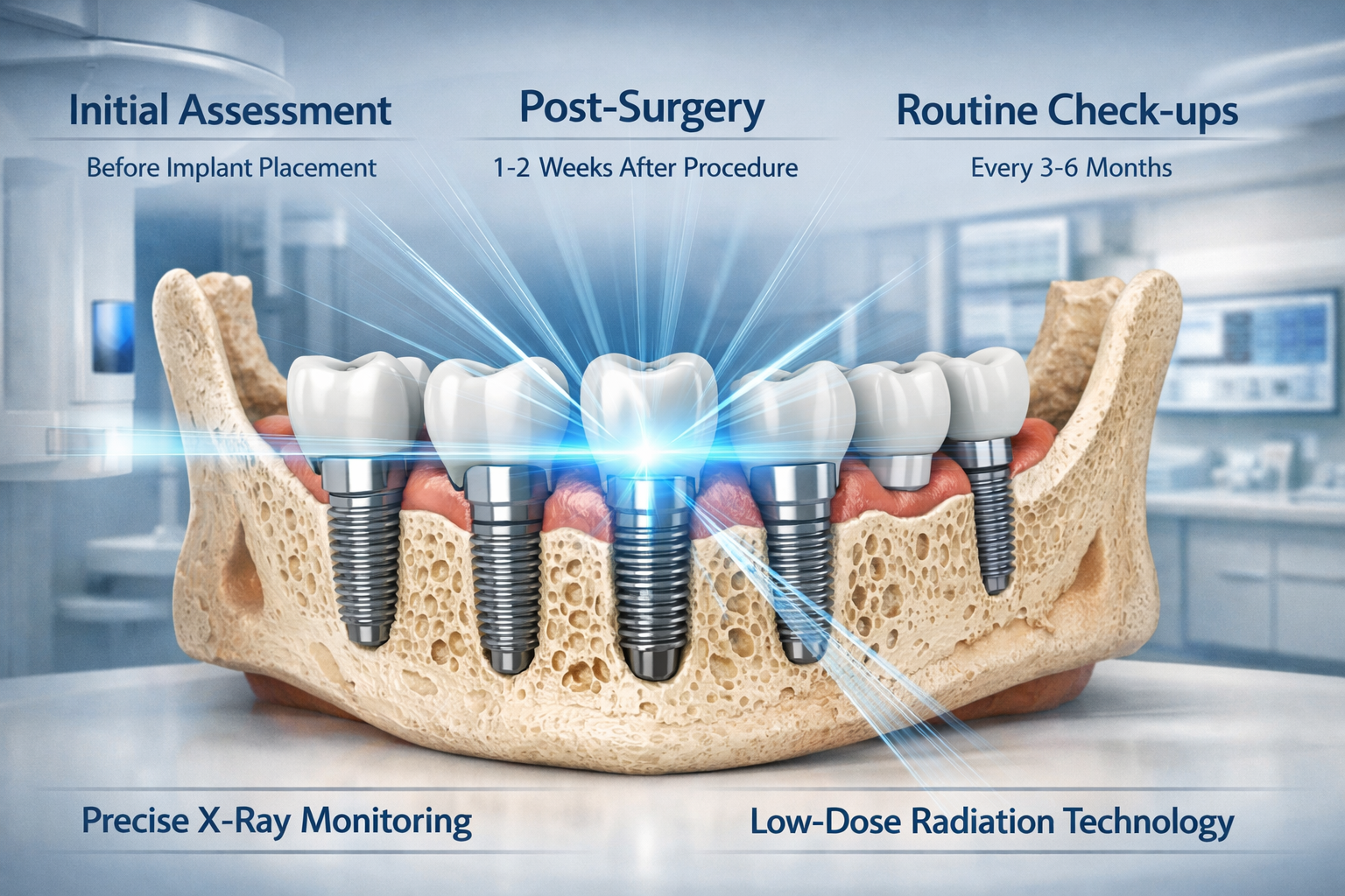
Immediately Post-Placement: After the implant is successfully placed, another X-ray is typically taken. This serves as a baseline image, documenting the implant’s position immediately after surgery. This initial X-ray is crucial for comparison with all future images, allowing your dentist to track any changes. This is part of the extensive complete dental implant recovery timeline and aftercare.
Establishing Your Dental Implant X-Ray Frequency: Post-Healing and Long-Term Maintenance
Once your dental implant has been successfully placed and the initial healing period is complete (which typically takes several months for osseointegration), the dental implant x-ray frequency shifts to a maintenance schedule. This phase is crucial for ensuring the implant’s long-term health and preventing complications.
The Standard Maintenance Schedule for Dental Implant X-Ray Frequency
For most healthy patients with stable implants and no specific risk factors, a typical maintenance schedule involves X-rays every 1 to 3 years [2]. This frequency is a balance between monitoring the implant’s health effectively and minimizing radiation exposure.
Here’s a breakdown:
- Year 1 Post-Restoration: A follow-up X-ray is commonly taken within the first year after the final crown or prosthesis is attached to the implant. This confirms that the implant-supported restoration is stable and that the surrounding bone is healthy after the loading period.
- Annual to Biennial Check-ups: For patients with excellent oral hygiene, no signs of complications, and stable bone levels, your dentist might recommend X-rays every two to three years as part of your comprehensive annual check-up.
- More Frequent Monitoring (Annually): If there are specific concerns, such as a history of periodontal disease, signs of early bone loss around other teeth, or if the implant is part of a complex full-arch restoration (like All-on-4 or All-on-6 implants), annual X-rays may be advised.
Factors Influencing Dental Implant X-Ray Frequency
The “standard” schedule is a guideline. Your individual dental implant x-ray frequency will be tailored by your dentist based on several key factors:
1. Your Overall Oral Health History
- History of Periodontal Disease: Patients with a history of gum disease are at higher risk for peri-implantitis (implant gum disease). More frequent X-rays (e.g., annually) may be necessary to monitor the bone supporting the implant.
- Bone Loss: If you’ve experienced significant 90 percent bone loss in teeth or required extensive bone grafting, your dentist might want to keep a closer eye on your implant sites.
2. Number and Type of Implants
- Single Implant vs. Multiple Implants/Full Arch: A single implant might require less frequent comprehensive imaging than a full arch of implants supporting a bridge (like All-on-4 dental implants). Full-arch restorations often involve more complex biomechanics and require careful monitoring of all implant sites.
- Immediate Load Implants: While effective, implants loaded immediately after placement might require closer initial monitoring compared to those that undergo a longer healing period before restoration.
3. Signs or Symptoms of Complications
If you experience any of the following, your dentist will likely recommend an immediate X-ray, regardless of your last scheduled one:
- Pain or discomfort around the implant.
- Swelling, redness, or pus around the gum line.
- A feeling of looseness in the crown or implant.
- Difficulty chewing or biting.
- Receding gums around the implant.
These symptoms could indicate peri-implantitis, an infection, or a mechanical issue. An X-ray is vital for diagnosis.
4. Systemic Health Conditions
Certain systemic conditions can impact bone health and healing, influencing the need for more frequent checks:
- Diabetes: Poorly controlled diabetes can affect healing and increase the risk of infections.
- Osteoporosis: While not an absolute contraindication, osteoporosis can affect bone density and may warrant closer monitoring.
- Smoking: Smokers have a significantly higher risk of implant failure and peri-implantitis, necessitating more vigilant X-ray monitoring.
5. Type of X-Ray Taken
Different X-ray types provide different levels of detail, which can influence how often they are used:
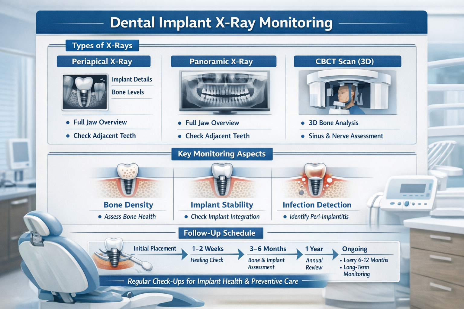
- Periapical X-rays: These highly detailed images focus on one or two teeth and the surrounding bone, providing a clear view of the implant’s apex and adjacent bone. They are excellent for localized monitoring and detecting early bone loss.
- Panoramic X-rays: These provide a broader view of the entire mouth, including both jaws, sinuses, and jaw joints. They are useful for screening and assessing the overall health of multiple implants and surrounding structures, but offer less detail than periapicals for individual implants.
- CBCT (Cone-Beam Computed Tomography) Scans: These 3D images offer the most comprehensive view, crucial for pre-surgical planning and diagnosing complex issues. However, due to higher radiation doses compared to 2D X-rays, they are typically reserved for specific diagnostic needs and not used for routine follow-up unless absolutely necessary.
| X-Ray Type | Primary Use | Typical Frequency (for Implants) | Detail Level | Radiation Dose |
|---|---|---|---|---|
| Periapical | Detailed view of single implant, bone level, and surrounding structures | As needed, or part of routine | High | Low |
| Panoramic | Overall view of jaws, sinuses, multiple implants, general screening | Every 1-3 years (routine) | Medium | Low-Medium |
| CBCT | 3D assessment for complex planning, diagnosis of specific issues | Pre-surgery, or for specific diagnosis | Very High | Medium-High |
My Philosophy on Dental Implant X-Ray Frequency
As an expert in this field, I always prioritize both diagnostic accuracy and patient safety. My approach to dental implant x-ray frequency is rooted in evidence-based dentistry and personalized care.
I advocate for a balanced approach:
- Initial Baseline: A comprehensive set of images post-placement is non-negotiable.
- First Year Follow-up: A critical checkpoint to ensure osseointegration and restoration stability.
- Personalized Routine: Subsequent frequency is always dictated by the individual patient’s risk profile, oral hygiene, and any emerging concerns.
- “As Needed” for Symptoms: If a patient presents with any discomfort or signs of trouble, an immediate X-ray is the fastest and most effective way to diagnose the problem.
- Lowest Effective Dose: We always use the most advanced digital X-ray technology available in 2026, which significantly reduces radiation exposure while providing superior image quality.
It’s important to remember that the benefits of detecting potential implant issues early, which can prevent more invasive and costly treatments, far outweigh the minimal risks associated with modern dental X-rays. For those considering dental implants, understanding the necessary follow-up care is crucial. You can begin your journey with a dental implant consultation to discuss these aspects.
Reducing Your Need for Emergency X-rays
While regular X-rays are crucial, maintaining excellent oral hygiene can help prevent many issues that might necessitate an unscheduled X-ray.
Here are my top tips:
- Brush and Floss Religiously: Treat your implants like natural teeth. Use a soft-bristled brush and floss daily, paying special attention to the areas around your implants. Many find a water flosser effective for cleaning around implants, as highlighted in best water flosser for dental implants.
- Regular Professional Cleanings: Beyond your dentist, a dental hygienist specializing in implant care can meticulously clean around your implants without causing damage. Learn more about dental implant cleaning cost to budget for this essential care.
- Avoid Hard Foods: While implants are strong, excessive force on very hard or sticky foods can strain them or the surrounding bone. Check out our guide on 50 soft foods after dental implant for safe choices.
- Quit Smoking: Smoking dramatically increases the risk of peri-implantitis and implant failure.
- Manage Systemic Diseases: Keep conditions like diabetes under control, as they can impact oral health.
By following these guidelines, you can contribute significantly to the long-term success of your implants and potentially reduce the frequency of urgent diagnostic X-rays.
Conclusion
Understanding your dental implant x-ray frequency is a critical aspect of being an informed dental implant patient. These seemingly simple diagnostic tools are the unsung heroes of long-term implant success, offering an invaluable window into the invisible health of your bone and implant integration. From precise pre-surgical planning in 2026 to ongoing vigilant monitoring for complications like peri-implantitis, X-rays play a non-negotiable role.
Remember, the specific frequency of your dental implant X-rays will always be a personalized decision made by your dentist, taking into account your unique oral health profile, the type and number of implants, and any existing risk factors. Always adhere to your dental professional’s recommendations, ask questions if you have concerns about radiation exposure (which is minimal with modern digital X-rays), and maintain impeccable oral hygiene. By doing so, you’re not just ensuring the health of your dental implants; you’re safeguarding your investment in a beautiful, functional smile for many years to come.
Actionable Next Steps:
- Schedule Your Regular Check-up: If you have dental implants, ensure you’re scheduling and attending your routine dental check-ups, which typically include a discussion about your X-ray needs.
- Discuss Your X-ray Schedule: During your next appointment, talk to your dentist about your specific dental implant x-ray frequency and why it’s recommended.
- Maintain Excellent Oral Hygiene: Continue to brush, floss, and potentially use a water flosser to keep the area around your implants clean and healthy.
- Report Any Concerns Immediately: Don’t wait until your next appointment if you notice pain, swelling, or any changes around your implants. Contact your dentist right away.
- Educate Yourself: Explore other resources on implant care on our site, such as insights into dental implant aftercare instructions or details on abutment placement to fully understand your treatment.
References
[1] Albrektsson, T., & Sennerby, L. (1990). State of the art in oral implants. Journal of Clinical Periodontology, 17(7), 415-420.
[2] American Academy of Periodontology. (2000). Position Paper: Dental Implants in Periodontal Therapy. Journal of Periodontology, 71(12), 1934-1942.
Dental Implant X-Ray Frequency Advisor (2026)
Your Oral Health & Implant Status
Risk Factors & Concerns
*This tool provides a general guideline based on common dental practices in 2026. It is NOT a substitute for professional medical advice. Always consult with your dental professional for personalized recommendations regarding your dental implant x-ray frequency and overall care.
Leave a Reply
Share your thoughts or ask a question about dental implants. Your email address will not be published.